I was the lab manager. I trained the PhDs; I do not have one.
Fifteen years in cell and gene therapy, bench to business. This page is the first half.
Two things it is here to prove. I ran an original research program that brought large grants into the lab. And I built the tooling and helped stand up the facility that other people's programs ran on. Both of those instincts show up again later, in software.
The sequence: Curt Freed's lab at CU Denver, in the embryonic stem cell era of Parkinson's cell therapy. Then Dennis Roop's, Ganna Bilousova's, and Igor Kogut's labs at the Gates Center for Regenerative Medicine, the center that became the Gates Institute (same labs, one name change), where the work was primarily iPSC-based.
On this page
The work below is in the order it matters, not the order it happened. My own program comes first, then the labs I helped run and what I contributed to them, then what I built for all of it to run on, then where I learned the basics.
If I did it, I say "I". If the lab did it, I say "we". If someone else's name is on the patent or the paper, their name is on it here.
- Engineering cells to find inflammation: my own program
- Dopamine neurons and Parkinson's disease: Freed lab, ESC era
- Harlequin ichthyosis and ABCA12: Roop lab
- iPSCs and RDEB: Gates, iPSC era
- Cellular rejuvenation
- Calling the next thing early
- Building the place
- Microscopy, high-throughput screening, and automation
- Where it started
- Published work: five papers
- What the bench taught me
Engineering cells to find inflammation
This is my thread. Mesenchymal stem cells (MSCs), modified with modified RNA, engineered first to home to inflamed tissue and then to change what they secrete once they arrive.
At Gates we were trying to reach the internal epithelium in skin and connective tissue disorders like recessive dystrophic epidermolysis bullosa (RDEB) and Ehlers-Danlos syndrome. MSCs are a good chassis for that. They can be isolated from bone marrow, from adipose tissue, from Wharton's jelly in umbilical cord, and they can be derived from induced pluripotent stem cells. They do not express major histocompatibility complex class II, which makes an off-the-shelf allogeneic product plausible instead of a per-patient build. And they damp inflammation and improve wound healing largely through what they release: exosomes carrying peptide and RNA cargo.
The problem is delivery. Infuse MSCs intravenously and they engraft poorly. They do not reliably leave the circulation for the tissue that needs them.
The grappling hook
Many groups had attempted a version of this in hematopoietic stem cells. The mechanism is glycosyltransferase-programmed stereosubstitution (GPS), which adds a sugar to CD44 and converts it into HCELL, the hematopoietic cell E- and L-selectin ligand. The fucosyltransferases FUT6 and FUT7 do that glycosylation efficiently.
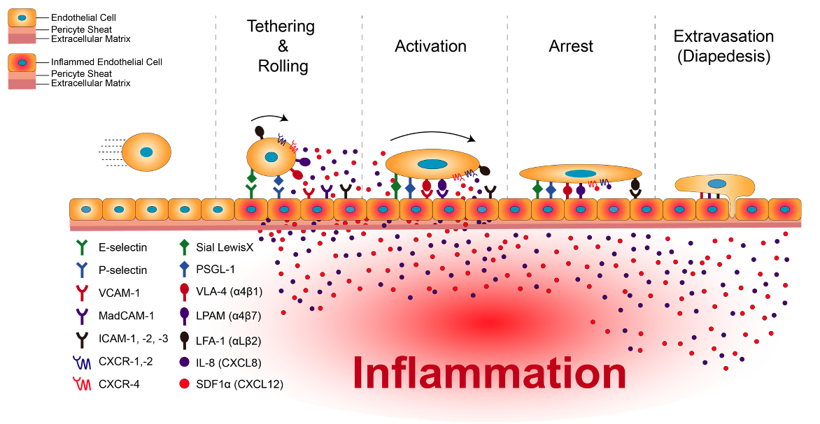
HCELL works like a grappling hook. When tissue is inflamed, the endothelium lining nearby vessels puts E- and L-selectin on its surface. A cell carrying HCELL catches on those selectins, slows down, tethers, rolls, arrests, and then extravasates, leaving the bloodstream to go to work.
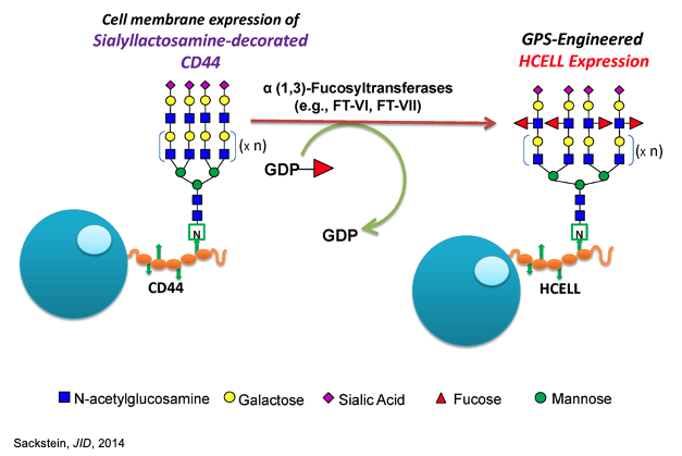
Those groups did the glycosylation to the cell, from outside. Our lab was an RNA lab, and held patents on RNA reprogramming and on non-integrating gene correction, so we asked a different question: what if we transfected the MSC with FUT6 RNA and let it glycosylate its own CD44?
The modRNA toolkit
Naked RNA is a poor drug. Cells read it as a virus: the innate sensors fire, translation shuts down, and the cell you were trying to help gets sick instead. So the RNA has to be built, not just transcribed.
That build is a set of specific choices, each with a job:
- Modified nucleotides in place of the standard ones, so the innate immune sensors do not recognize the transcript as foreign.
- Optimized poly-A tails, which set how long the message survives before it is degraded.
- ARCA and methylation caps. An anti-reverse cap analog forces the cap onto the transcript in the correct orientation, so ribosomes can load it instead of wasting it, and methylating the cap makes it read as the cell's own message rather than as something to attack.
- Extensive molecular cloning upstream of all of it, because every construct, tag, and untranslated region has to be built and tested before any of the above matters.
Get those right and you have a transient, non-integrating way to make a cell express whatever you want, for a defined window, without touching its genome.
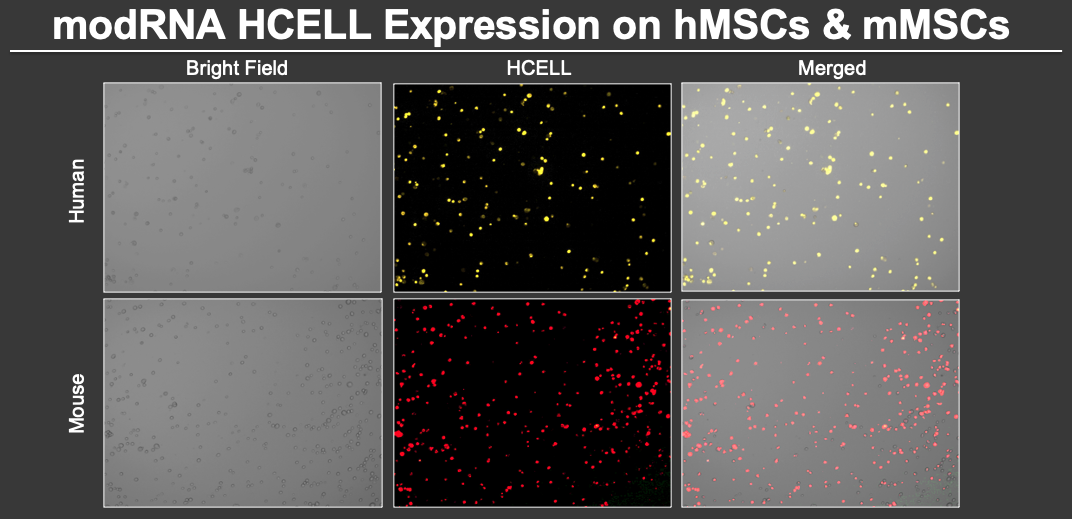
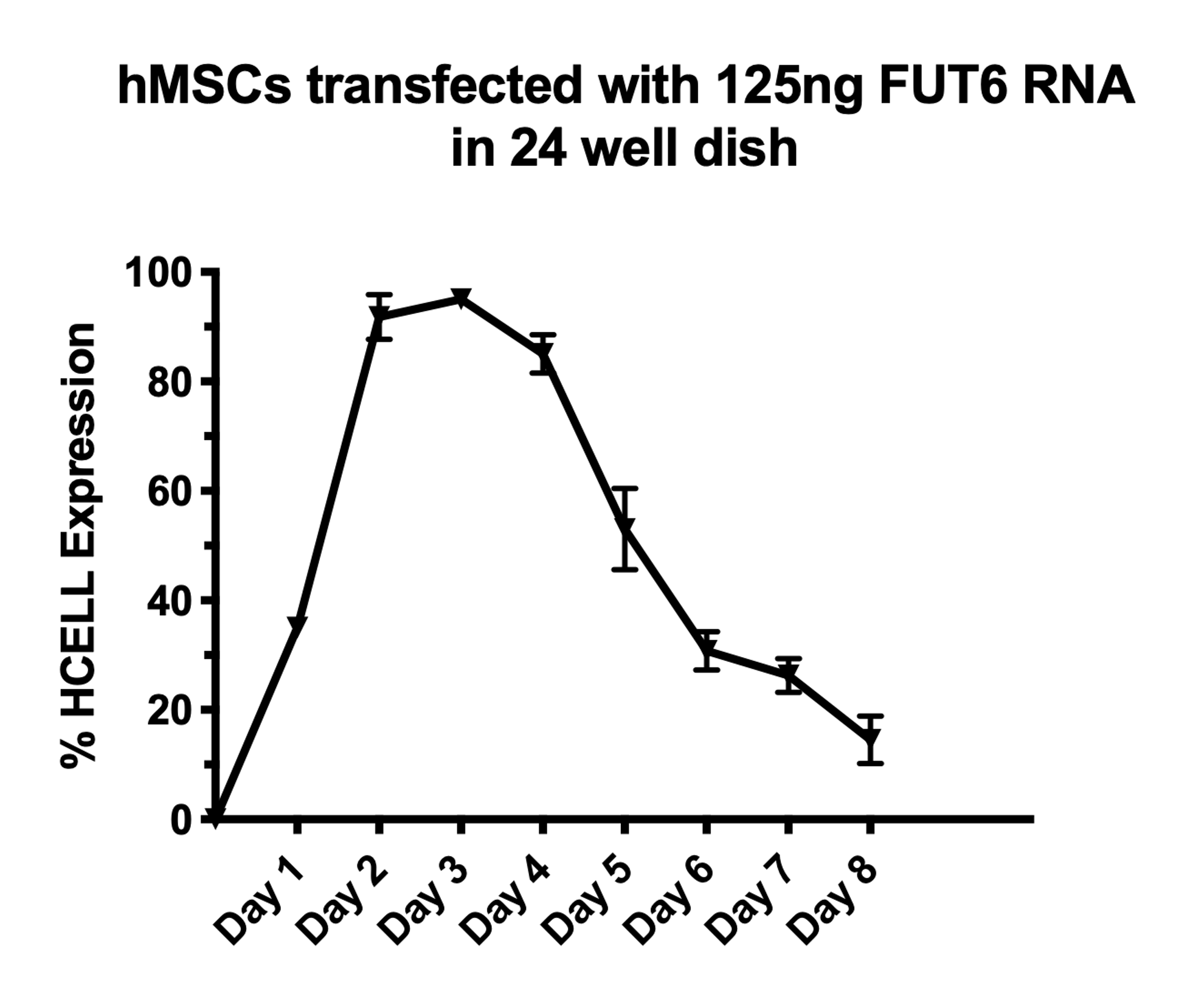
It worked. FUT6 modRNA induced HCELL expression on both human and mouse MSCs. Expression peaked at 48 hours and stayed high for another 48 hours after that: a four-day expression window from a single transfection, and then the cell goes back to being itself.
Does it actually home?
Surface expression in a dish is not homing. So I went and looked.
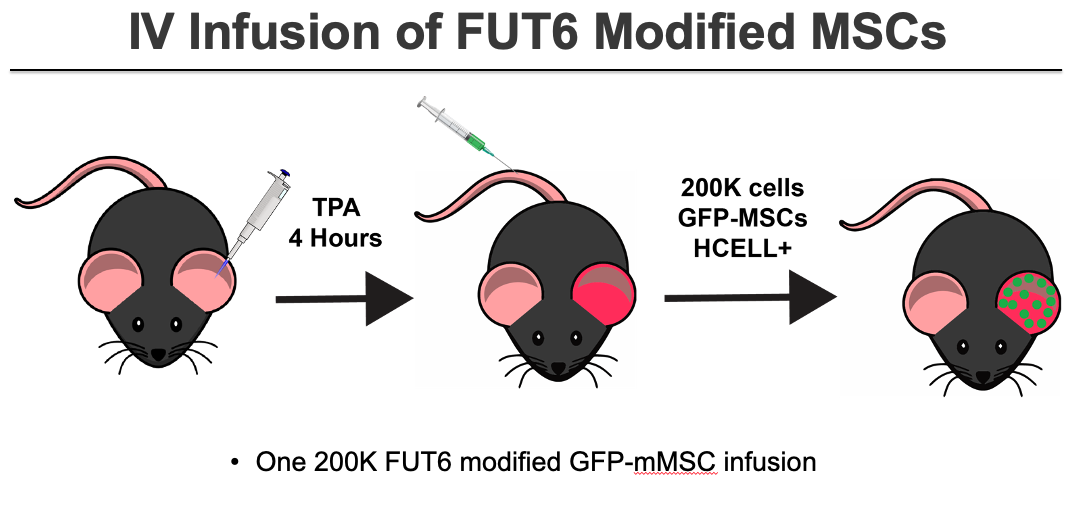
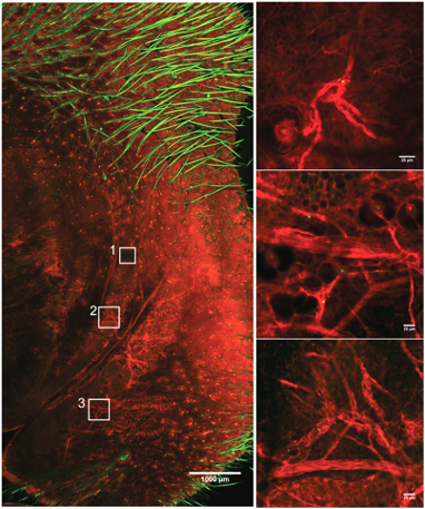
I injected TPA, an irritant, subcutaneously into a mouse engineered to express the red fluorescent protein tdTomato. Then I delivered HCELL-modified, GFP-labeled MSCs by tail vein. Then I put the animal's ear under a point-scanning confocal microscope in an intravital imaging chamber I built, and watched for green cells in red tissue.
Within hours of the infusion, green cells that had entered at the tail were in the ear. One of them left the bloodstream while I was recording it.
The other advantage of intravital imaging is that the mouse survives it. The same animal can be imaged again and again, so engraftment can be followed as a time course in one animal rather than as a series of endpoints across many.
Then, what the cell says when it gets there
Homing is only logistics; the therapy is what the cell secretes once it arrives.
The second half of the program used the same modRNA chassis with a different payload, engineering the paracrine signaling of the cell so that what it releases into the tissue is chosen rather than inherited. Same delivery vehicle, different cargo.
Homing and paracrine engineering were one program, not two. This work brought large grants into the lab.
Dopamine neurons and Parkinson's disease
Curt Freed's lab, the embryonic stem cell era.
Freed's lab worked Parkinson's from two directions: cell replacement with embryonic stem cells, and pharmacological activation of the neuroprotective pathways that defend cells against oxidative stress.
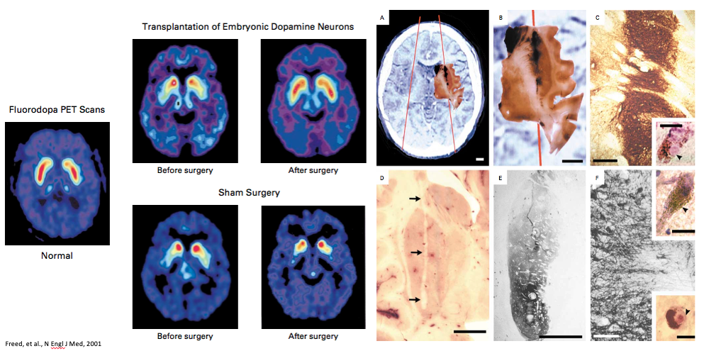
The lab's foundational result predates me. In the 1990s, Drs. Curt Freed and Robert Breeze performed the first-ever double-blind neurotransplantation surgery: four injections of human fetal ventral mesencephalic cells, in a trial of 40 patients aged 34 to 75 randomized to transplant or sham. The trial found that patients who had responded well to levodopa did well after the procedure, and patients with a historically poor levodopa response benefited less. That is Freed and Breeze's work, published in the New England Journal of Medicine, not mine.
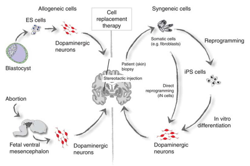
Fetal tissue carries obvious ethical constraints and an obvious supply constraint. Induced pluripotent stem cells removed both in principle: take an adult cell, reprogram it to pluripotency, and you have an unlimited source. In practice the constraint moved rather than disappeared. Anything left undifferentiated in the graft is a tumor risk, so purity becomes the gating question. Either you differentiate completely, or you find a marker that lets you sort the cells you want from the cells you do not. Kikuchi et al. took the second route: dopaminergic progenitors sorted on the floor-plate marker CORIN, transplanted into a primate model of Parkinson's, survived, extended neurites into the host striatum, improved spontaneous movement, and formed no tumors for at least two years. That result is the argument for the sorting step, not a reason to skip it.
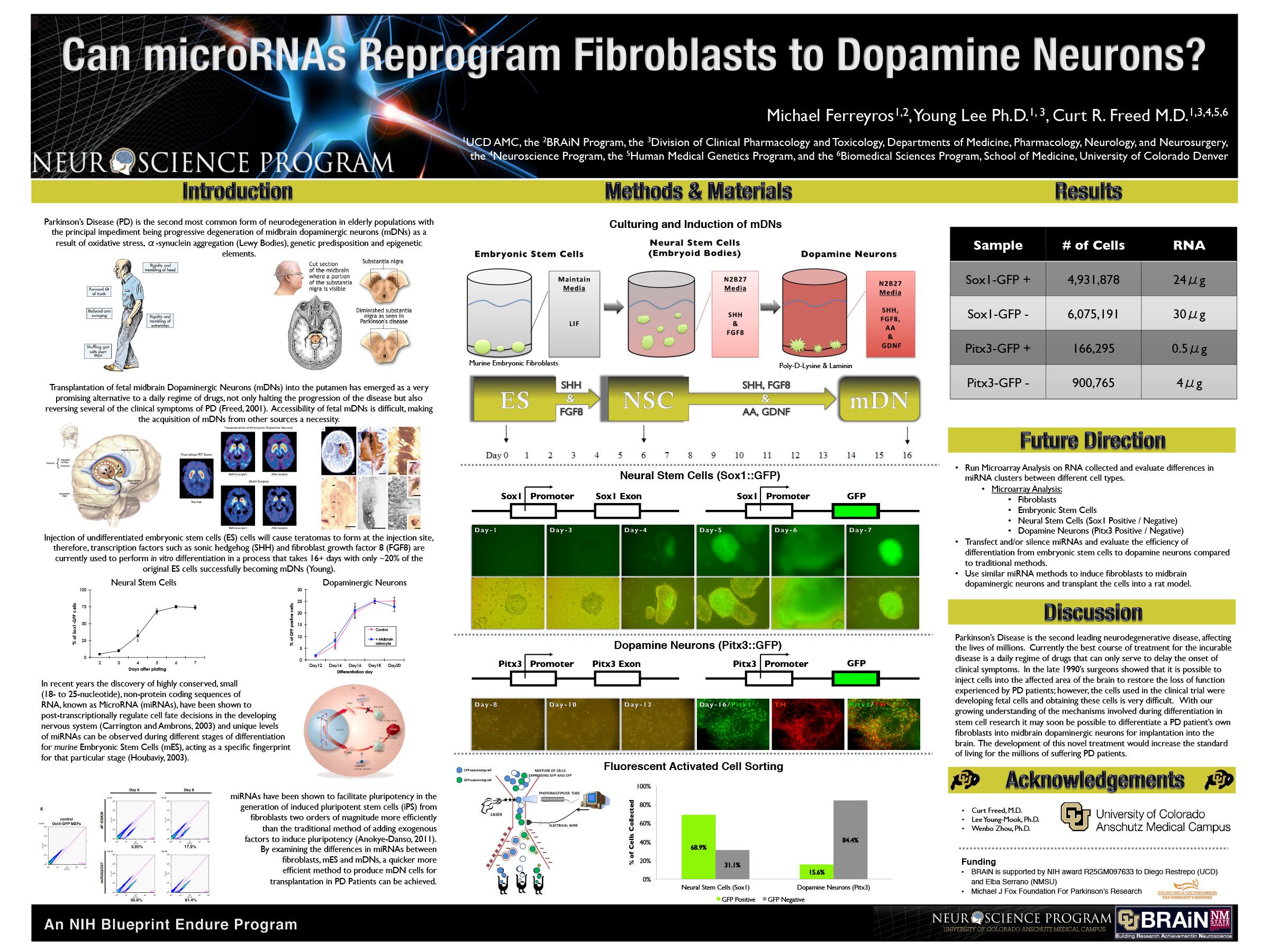
As a participant in the NIH Blueprint ENDURE program, I worked with Dr. Curt Freed and Dr. Wenbo Zhou on the feasibility side of a cell replacement therapy. Reports on boosting reprogramming efficiency with microRNA were coming out steadily, and we asked whether you could skip pluripotency entirely: transdifferentiate fibroblasts straight into dopamine neurons with a microRNA cocktail. The approach was empirical: isolate microRNAs, sequence them to catch both known and unknown species, normalize pseudo-reads with the CU Anschutz biostatistics department. Then take the top 16 significantly up- and down-regulated species forward, and use them to drive conversion of human ES and iPS cells toward dopamine neurons. The Pitx3-positive versus Pitx3-negative contrast is what made the screen readable.
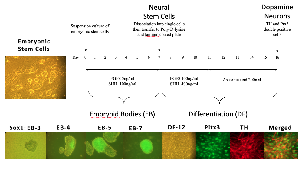
The run, in order:
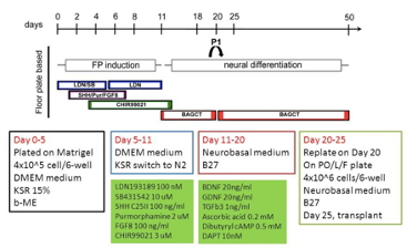
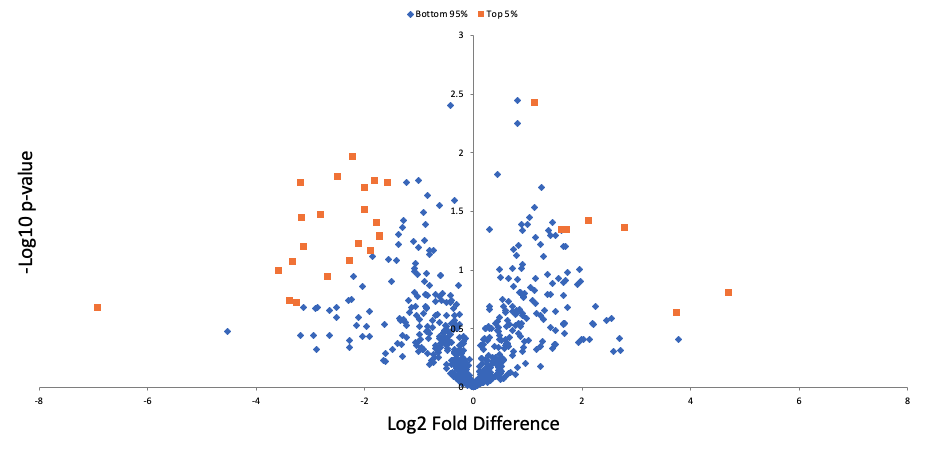
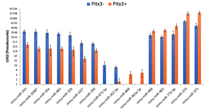
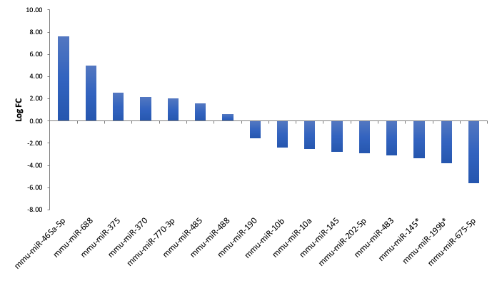
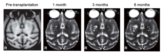
The in vivo model is a unilateral 6-OHDA injection. 6-OHDA looks enough like dopamine to be taken up by dopamine neurons and different enough to kill them, so one injection wipes out the dopaminergic population in the substantia nigra on one side. Those A9 neurons have their cell bodies in the nigra and project axons to the striatum, so when they die the striatum stops receiving dopamine, and challenge with apomorphine makes the animal turn in circles. Transplant differentiated cells into the striatum and you score functional recovery by counting rotations.
Harlequin ichthyosis and ABCA12
Dennis Roop's lab.
Roop's lab works on heritable blistering skin and connective tissue disorders. On this project I picked up previous post-doctoral students' unfinished work and carried it through to publication.
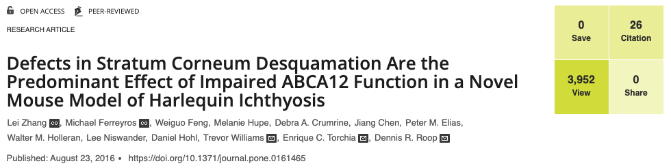
Harlequin ichthyosis is a severe, potentially lethal neonatal skin disease caused by mutations in ABCA12. The lab identified a novel mutation in the mouse Abca12 gene that produces a comparable disease in the animal, which makes it a usable model for testing treatments.
The mechanism is transport. ABCA12 delivers glucosylceramides and corneodesmosin to where the skin barrier is assembled. Lose it and the lipid lamellae in the epidermis never form. The mutant mice also had reduced kallikrein 5 and kallikrein 7 (the proteases required for normal shedding) so the barrier fails to build and the outer layer fails to come off. One transporter, both halves of the phenotype.
iPSCs and RDEB
Gates, the iPSC era.
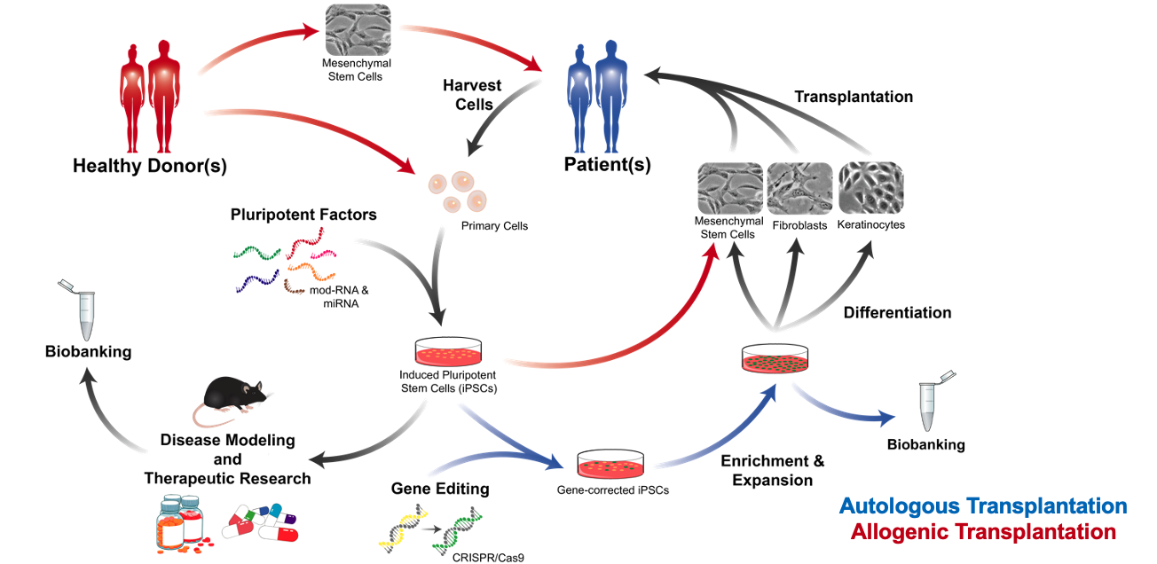
Gates-era work was primarily iPSC-based. The reason, as I understood it, is arithmetic. In a recessive genetic skin disease, a corrected cell is worth more than a healthy donor cell, because it is the patient's own: no immune mismatch, no lifelong immunosuppression. But you cannot correct and expand enough cells at the tissue level. You can at the pluripotent level.
Drs. Bilousova and Kogut hold the patents that make that route practical: a highly effective RNA reprogramming technique, and a single-step iPSC gene correction protocol built on a reverse-transcriptase Cas system (prime editing) that raises editing efficiency while keeping indels low. Those are their inventions. I helped run the labs where they were used, and trained the people who used them.
The sequence those two patents unlock is clean. Take a patient's cells. Reprogram them with RNA, non-integrating, so nothing is left behind in the genome. Correct the mutation in the pluripotent state, where a single edited clone can be expanded without limit and sequenced before anything is put back into a person. Then differentiate, and testing what you differentiated needs skin to put it in, which is the multilayer human skin reconstruction model I co-authored the protocol for.
MSCs can be derived from iPSCs; I co-authored the protocol for that differentiation. So a corrected line could in principle feed the delivery work above: a patient-matched, gene-corrected MSC that homes where it is needed. To be clear about what that sentence is: the route the two threads point at, not a result. Nobody has handed me a patient-matched homing MSC. It is the reason the two programs belonged in one building, and it is unfinished.
Cellular rejuvenation
Drs. Bilousova and Kogut developed a rejuvenation cocktail that uses CRISPR activation to upregulate factors that lengthen telomeres and stabilize metabolic function. Theirs. What I can show is the readout.
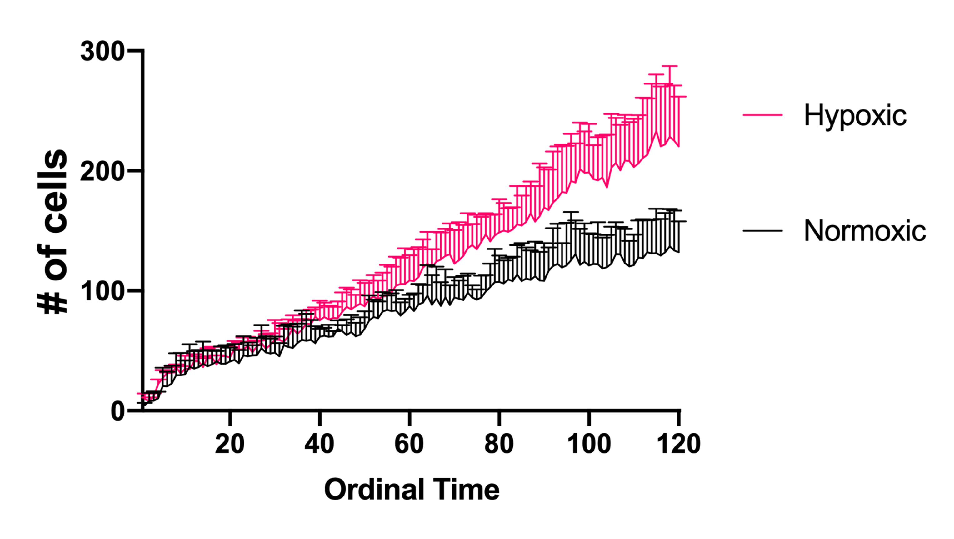
That readout is behavioral, not molecular, and that is the point. A marker panel tells you what a cell is. A chemotaxis assay tells you what it does. The first clip is the baseline the cocktail has to beat, not the cocktail working: young donor cells and old donor cells run toward hepatocyte growth factor, imaged side by side on the same time base. The second clip is a run of the rejuvenation assay itself.
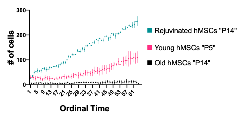
I am showing the footage and not a number. The imaging exists; the quantified, peer-reviewed result is the lab's to publish, and I will not put an effect size on a website that has not been through review.
Calling the next thing early
Some of what I contributed was not an experiment. It was an argument about where to point.
I pushed the lab into exosomes, lipid nanoparticles, and the senescence-associated secretory phenotype years before any of the three were fashionable. The reasoning was the same in each case, and it followed directly from the MSC work: if the therapeutic effect of these cells travels in released vesicles, then the vesicle is the drug and you should study it directly. If a transient, non-integrating RNA payload is the mechanism, then the packaging that delivers it into a patient is not a detail, it is the product. And if aging cells signal differently, then the secretome you are trying to engineer changes with donor age, which makes senescence a variable in your therapy whether or not you measure it.
I make the same kind of call now on the business side, where the argument is about AI instead of vesicles.
Building the place
I helped stand up the institute: equipment purchasing, facilities, the physical build-out. The photographs below are of what that build-out produced. Same labs, under the name they carry now.
The decision I would defend hardest is the one about direction. The cGMP facility was mirrored to the research labs, not the other way around. Research picked its equipment for scientific reasons (what a discovery scientist actually needs to see and do) and the GMP suite was then built to match it. That ordering is deliberate. If the manufacturing floor dictates the bench, discovery gets constrained by a process that does not exist yet. If the bench dictates the floor, a protocol that works at research scale crosses into GMP with fewer translations, fewer requalifications, and fewer arguments about why the clinical result does not look like the paper.
Vendor selection followed the same logic. STEMCELL Technologies and Miltenyi were chosen with the clinic in mind from day one, because a reagent or an instrument you cannot follow into a regulated process is a wall you will hit later, at the worst possible moment.
The rest of the build-out was ordinary engineering with the same bias toward the person doing the work. Fifteen hypoxic incubators in the tissue culture suite run on house gas through a custom liquid-to-gas interchange built into the lab, a design that gave the culture specialists back their working space. The tissue culture suites are laid out around a sterile entry and a quarantine room where off-site samples wait for mycoplasma testing before they are allowed anywhere near a shared incubator. Samples are tracked in Freezerworks, and the Luminex in the lab runs SASP panels.
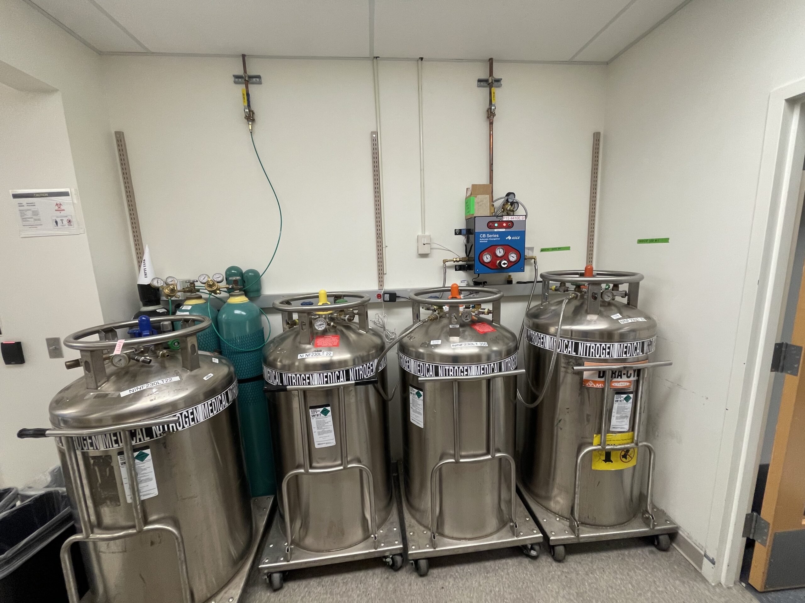
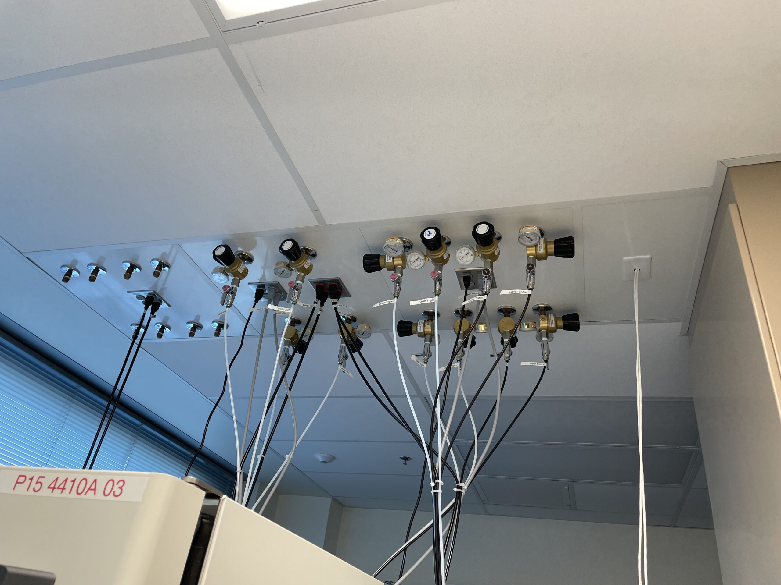
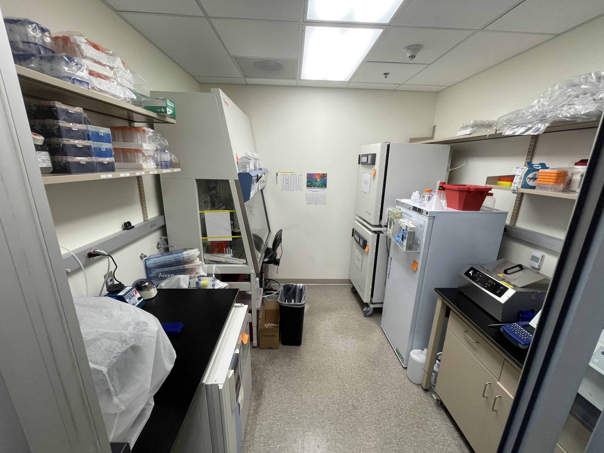
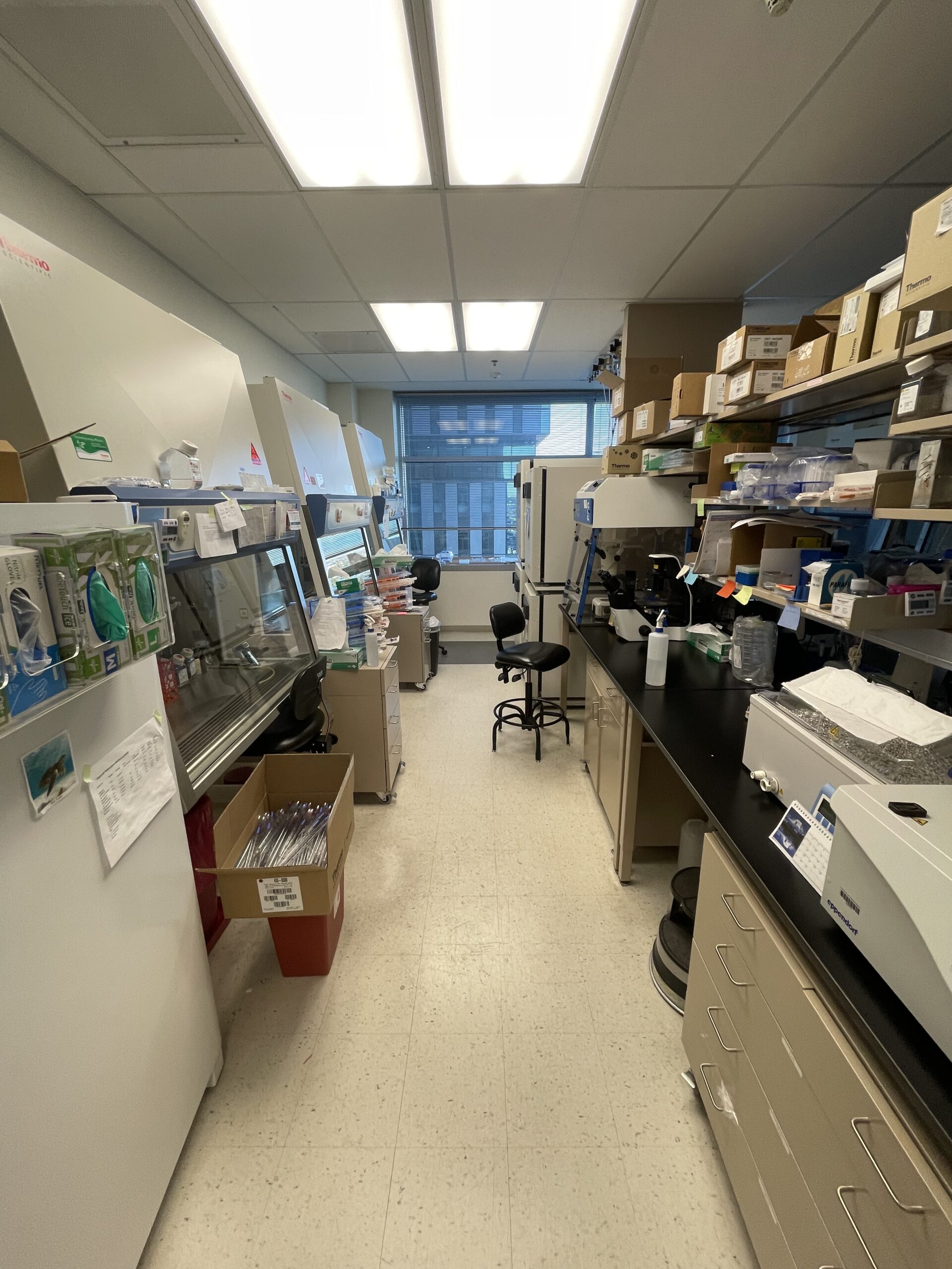
None of this is glamorous. All of it is why an experiment runs on the day someone decided to run it.
Microscopy, high-throughput screening, and automation
At Gates I built the assays that made the homing work measurable: a transendothelial migration assay adapted for high-throughput screening, two shear flow assays for in vitro homing efficiency, and the intravital tooling for in vivo homing described above.
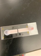
The shear flow work uses an ibidi pump system and µ-slides to put modified MSCs under physiological flow over immobilized protein. That confirmed the affinity for E- and L-selectin under conditions that resemble a vessel rather than a dish. Binding at zero shear tells you very little about a cell in circulation. To test an individual protein, I inserted a western blot membrane carrying the recombinant protein between the slide and the flowing cells, and watched whether the cells slowed on it.
Transendothelial migration, at plate scale
Once we could slow the cells down, the question became which modifications actually got them out of the vessel. Slowing on selectin is necessary but it is not the endpoint; extravasation is. That is what the transendothelial migration assay measures: cells crossing a living endothelial monolayer rather than sticking to it.
Run by hand it is a slow, one-condition-at-a-time experiment, and one condition at a time is how a screening question dies. So I rebuilt it for high-content analysis on a CellInsight CX7 with on-stage incubation: live cells held at temperature and CO2 on the instrument, imaged automatically across a full plate, and scored by image analysis instead of by eye. One run then answered what used to take weeks of separate experiments, and every condition was measured the same way rather than by whichever person happened to be at the scope.
That is the same instinct as everything on the work page, ten years earlier and in glassware: find the step that only scales with human hours, and take the human hours out of it.
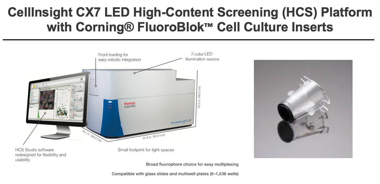
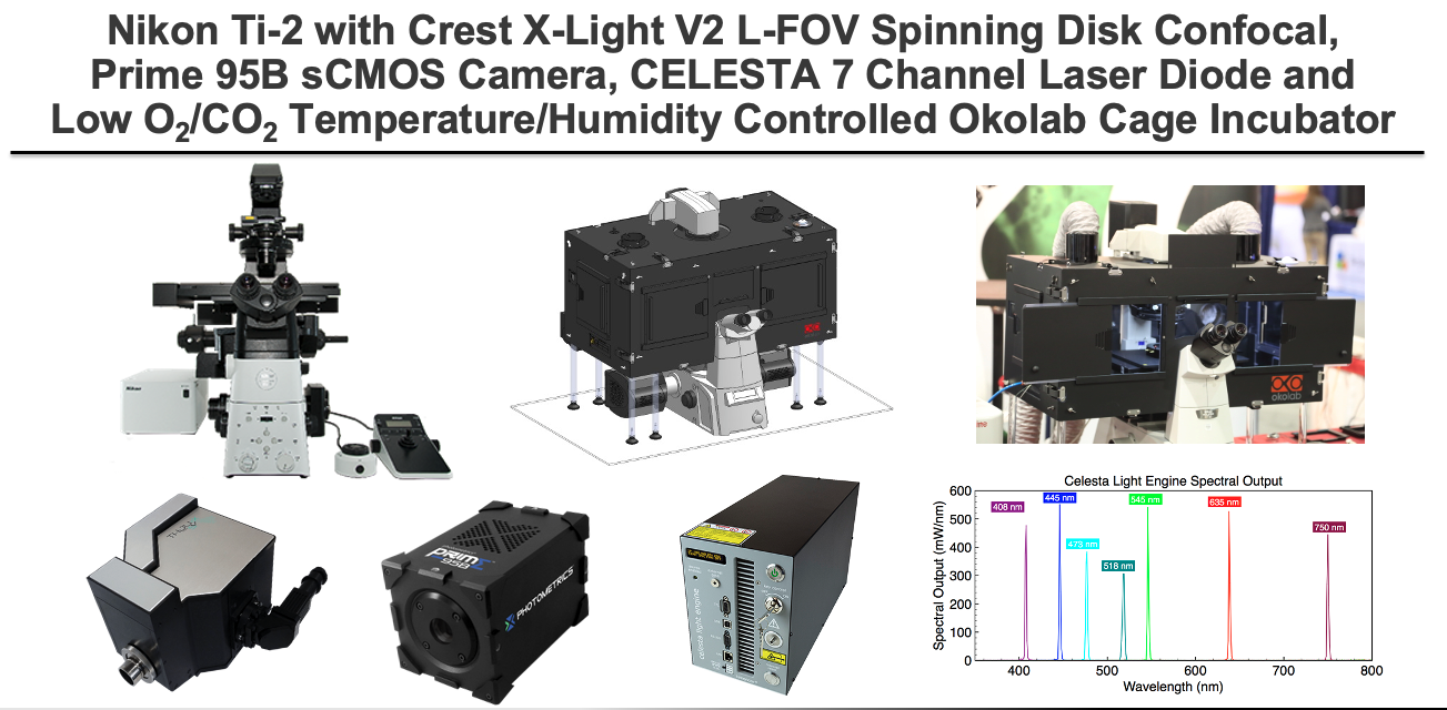
Donor funding paid for the imaging that made all of it possible, including the spinning disk confocal: a Nikon Ti2 with a CREST V2 spinning disk, Prime camera, seven-channel illumination, and Okolab stage-top incubation, which is what lets you image live cells for hours without killing them.
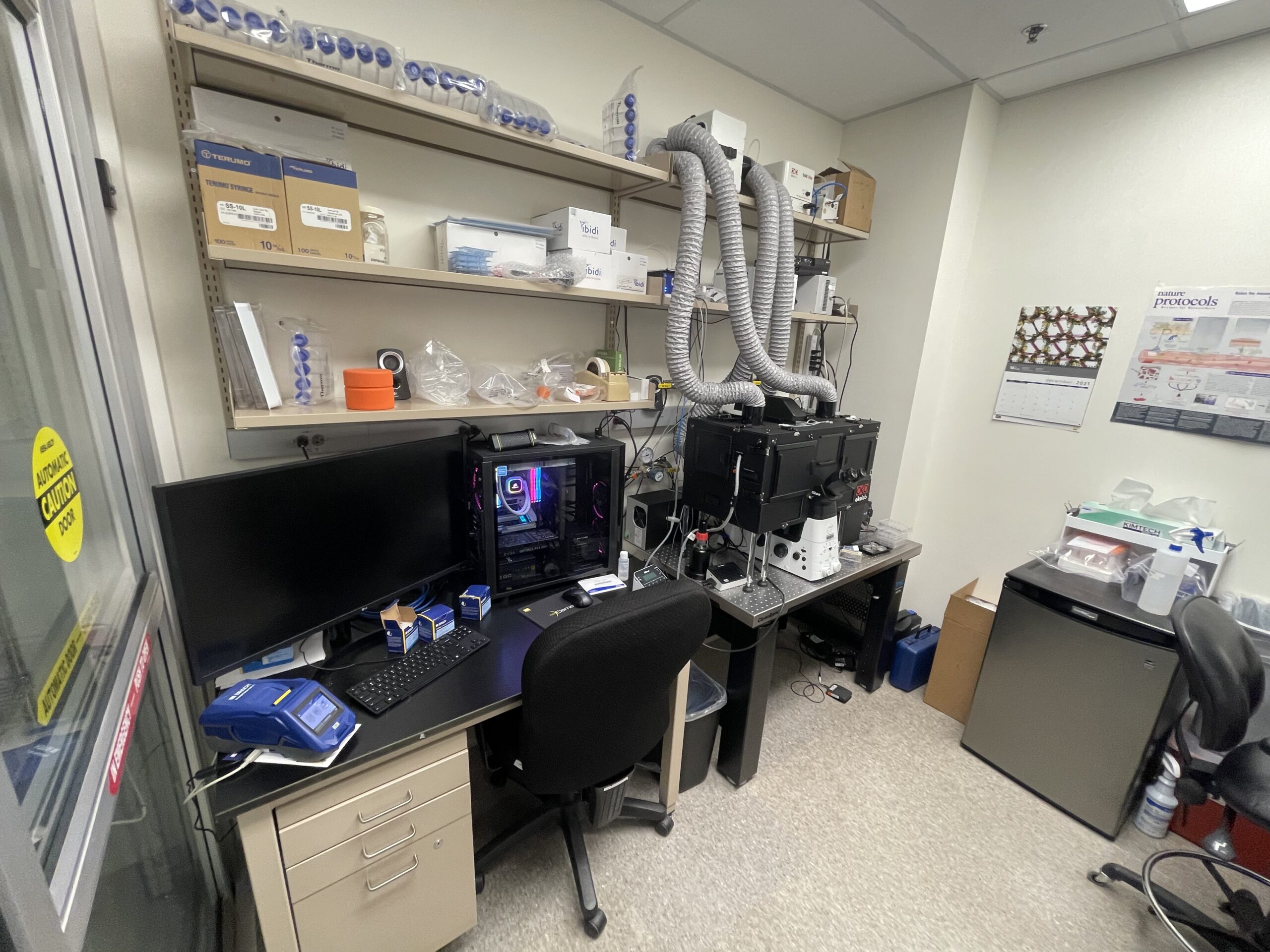
The through-line, and the reason this section belongs on a page about a bench career: every one of these was an automation. A homemade imaging chamber, a membrane trick, a plate reader protocol. I build the same things now, out of software.
Where it started
My first lab was Dr. Amanda Charlesworth's, on the post-transcriptional control that zygotic arrest proteins exert during fertilization and early embryogenesis.
In early development, the mother's stored maternal mRNAs are translated into the proteins the embryo needs, but that translation is timed, not automatic. Zar1 and Zar2 are part of the timing mechanism. Zar2 binds a translational control sequence in specific maternal mRNAs and regulates their translation; Zar1 does something similar by a less understood route. We showed that both bind the translational control sequence through a zinc finger in the C-terminal domain, and that Zar1 has the higher affinity for RNA. Similar function, likely different roles.
I am permanently grateful to those first labmates for teaching me the basics.
Published work
Five papers, one of them a preprint. Four appear above, in the sections they came out of.
- McGarvey SS, Ferreyros M, Kogut I, Bilousova G. Differentiating induced pluripotent stem cells toward mesenchymal stem/stromal cells. Methods in Molecular Biology, 2022. PMID 33772462
- Pavlova M, Balaiya V, Flores JC, Ferreyros M, Bush K, Hopkin A, Kogut I, Roop DR, Bilousova G. The development of an advanced model for multilayer human skin reconstruction in vivo. Bio-protocol, 2024. PMID 38268973
- Jayakumar RP, Sueoka T, Ferreyros M, Li BY, Madhav MS, Chen X, Knierim JJ, Cowan NJ. Synchronized path-integration recalibration and distinct landmark-control dynamics in head direction and CA1 place cells. bioRxiv, 2025. PMID 41279962. A preprint, not peer reviewed, and the outlier in this list: hippocampal electrophysiology rather than stem cells.
- Yamamoto TM, Cook JM, Kotter CV, Khat T, Silva KD, Ferreyros M, Holt JW, Knight JD, Charlesworth A. Zar1 represses translation in Xenopus oocytes and binds to the TCS in maternal mRNAs with different characteristics than Zar2. Biochimica et Biophysica Acta, 2013. PMID 23827238
- Zhang L, Ferreyros M, Feng W, Hupe M, Crumrine DA, Chen J, Elias PM, Holleran WM, Niswander L, Hohl D, Williams T, Torchia EC, Roop DR. Defects in stratum corneum desquamation are the predominant effect of impaired ABCA12 function in a novel mouse model of harlequin ichthyosis. PLoS One, 2016. PMID 27551807 Co-first author.
What the bench taught me
RNA is a control surface. You can tell a cell where to go and what to say, for a defined window, without editing its genome, and the hard part is never the idea; it is the poly-A tail and the cap and the clones underneath.
The other lesson took longer. None of this work reached a patient because it was good. It reached as far as it did because the equipment was bought for the right reason, the GMP suite matched the bench, the vendors could be followed into a regulated process, and someone did the grant management. Translational medicine is a team sport. I learned that at the bench, which is why I went and did the other half.
The science I still follow: longevity, skin rejuvenation, and personalized autoimmune-derived CAR-T.
That other half is the work, the data hubs and evidence tools and automations I build now, and the teaching, which is how it spreads past me.